


-
摘要:
为了降低患者的辐射风险,低剂量CT(LDCT)广泛用于临床诊断,但辐射剂量的减少在重建的LDCT图像中引入了斑点噪声和条纹伪影。为了提高LDCT图像的质量,提出了一种基于可变阶变分模型的后处理技术。所提出的变分模型使用边缘指示器控制变分阶数,根据图像的特征在一阶全变分(TV)正则项和二阶有界Hessian(BH)正则项之间交替变换。采用基于快速傅里叶变换(FFT)的分裂Bregman算法求解所提出的变分模型。该模型在保留高剂量CT(HDCT)图像相应结构的同时,有效抑制了斑点噪声和条纹伪影。重建的图像和实验数据表明,所提出的变分模型比现有的先进模型具有更好的质量。
-
关键词:
- 低剂量CT (LDCT) /
- 图像降噪 /
- 边缘指示器 /
- 全变分(TV) /
- 有界Hessian (BH) /
- 快速傅里叶变换(FFT) /
- 分裂Bregman算法
Abstract:Low-dose CT (LDCT) is widely used for clinical diagnosis to reduce radiation risk to patients. However, the radiation dose reduction introduces mottle noise and streak artifacts into the reconstructed LDCT images. In this paper, a post-processing technique is proposed based on variable order variational model to improve the LDCT image quality. The proposed variational model employs the edge indicator to control the order of variation, which can alternate between the first order total variation (TV) regularizer and second order bounded Hessian(BH) regularizer based on the image feature. Moreover, the proposed model is solved by split Bregman algorithm based on fast Fourier transform (FFT). The proposed model effectively suppresses mottle noise and streak artifacts, meanwhile preserving structure in reference to high-dose CT (HDCT) images. The reconstructed images and experimental data indicate that the proposed model has better quality than some existing state-of-the-art models.
-

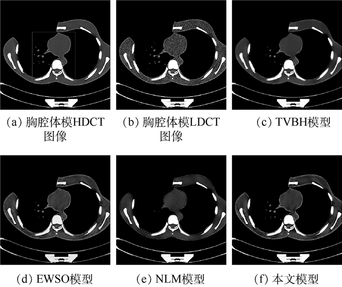
图 1 真实胸腔体模LDCT图像降噪结果的视觉比较
Figure 1. Visual comparison of denoising results on LDCT image of actual thoracic phantom

图 2 真实胸腔体模局部放大图的视觉比较
Figure 2. Visual comparison of denoising results on the local enlarged drawing by the squares in Fig. 1(a).

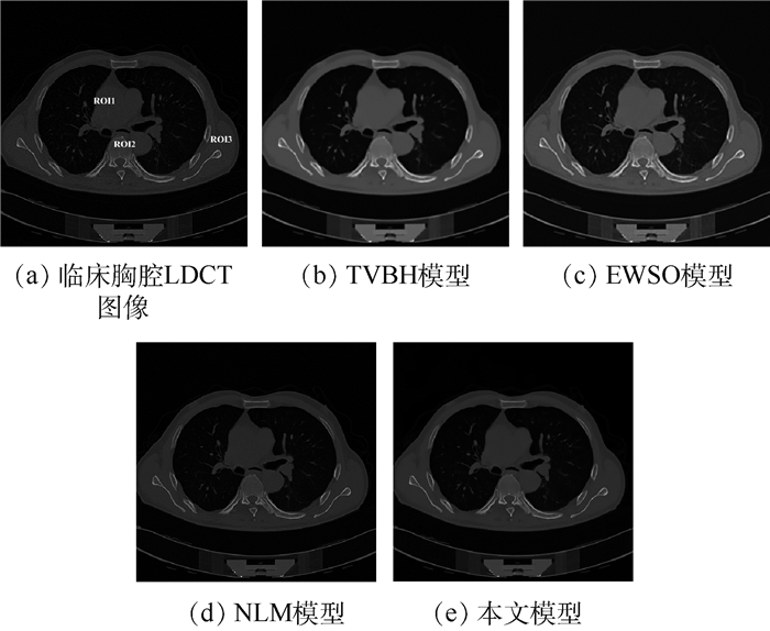
图 3 临床胸腔LDCT图像降噪结果的视觉比较
Figure 3. Visual comparison of denoising results on LDCT image of clinical thoracic

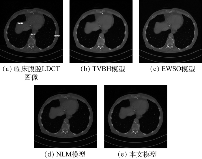
图 4 临床腹腔LDCT图像降噪结果的视觉比较
Figure 4. Visual comparison of denoising results on LDCT image of clinical abdominal

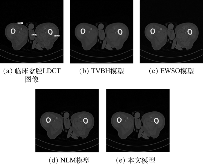
图 5 临床盆腔LDCT图像降噪结果的视觉比较
Figure 5. Visual comparison of denoising results on LDCT image of clinical pelvic
表 1 实际胸腔体模的定量比较
Table 1. Quantified comparison of actual thoracic phantom
降噪模型 
PSNR/dB MSSIM 除噪时间/s TVBH模型 25.259 3 0.886 4 19.45 EWSO模型 24.213 9 0.914 9 10.83 NLM模型 26.664 1 0.908 8 379.78 本文模型 27.205 1 0.926 6 27.88  下载: 导出CSV
表 2 临床数据的SNR值比较
Table 2. Comparison of SNR values of clinical data
LDCT图像及降噪模型 


ROI1 ROI2 ROI3 ROI1 ROI2 ROI3 ROI1 ROI2 ROI3 LDCT图像 0.529 0 0.049 7 0.093 7 0.073 3 0.275 6 0.120 0 0.067 0 1.201 4 0.069 9 TVBH模型 9.306 3 0.054 7 0.098 3 0.165 0 0.697 8 0.209 4 0.341 7 2.421 4 0.078 0 EWSO模型 14.386 6 0.054 6 0.099 2 0.093 5 0.485 6 0.162 4 0.108 5 1.320 2 0.072 2 NLM模型 1.301 1 0.058 0 0.094 6 0.173 1 0.890 9 0.159 4 0.110 5 1.446 7 0.069 7 本文模型 26.270 6 0.360 8 2.099 4 0.875 0 1.288 4 1.129 7 0.641 6 3.049 5 0.093 1
下载: 导出CSV
-
[1] ZHU Y, ZHAO M, ZHAO Y, et al.Noise reduction with low dose CT data based on a modified ROF model[J].Optics Express, 2012, 20(16):17987-18004. doi: 10.1364/OE.20.017987 [2] CHEN Y, YIN X, SHI L, et al.Improving abdomen tumor low-dose CT images using a fast dictionary learning based processing[J].Physics in Medicine and Biology, 2013, 58(16):5803-5820. doi: 10.1088/0031-9155/58/16/5803 [3] ZHANG C, ZHANG T, LI M, et al.Low-dose CT reconstruction via L1 dictionary learning regularization using iteratively reweighted least-squares[J].BioMedical Engineering OnLine, 2016, 15(1):66. doi: 10.1186/s12938-016-0193-y [4] LEE D, LEE J, KIM H, et al.A feasibility study of low-dose single-scan dual-energy cone-beam CT in many-view under-sampling framework[J].IEEE Transactions on Medical Imaging, 2017, 36(12):2578-2587. doi: 10.1109/TMI.2017.2765760 [5] CHEN Y, LIU J, HU Y, et al.Discriminative feature representation:An effective postprocessing solution to low dose CT imaging[J].Physics in Medicine and Biology, 2017, 62(6):2103-2131. doi: 10.1088/1361-6560/aa5c24 [6] CHEN Y, LIU J, XIE L, et al.Discriminative prior-prior image constrained compressed sensing reconstruction for low-dose CT imaging[J].Scientific Reports, 2017, 7(1):13868. doi: 10.1038/s41598-017-13520-y [7] FRUSH D P, DONNELLY L F, ROSEN N S.Computed tomography and radiation risks:What pediatric health care providers should know[J].Pediatrics, 2003, 112(4):951-957. doi: 10.1542/peds.112.4.951 [8] BRENNER D J, HALL E J.Computed tomography-An increasing source of radiation exposure[J].New England Journal of Medicine, 2007, 357(22):2277-2284. doi: 10.1056/NEJMra072149 [9] CHEN Y, SHI L, YANG J, et al.Radiation dose reduction with dictionary learning based processing for head CT[J].Australasian Physical and Engineering Sciences in Medicine, 2014, 37(3):483-493. doi: 10.1007/s13246-014-0276-7 [10] YANG Q, YAN P, ZHANG Y, et al.Low dose CT image denoising using a generative adversarial network with wasserstein distance and perceptual loss[J].IEEE Transactions on Medical Imaging, 2018, 37(6):1348-1357. doi: 10.1109/TMI.2018.2827462 [11] LIU J, MA J, ZHANG Y, et al.Discriminative feature representation to improve projection data inconsistency for low dose CT imaging[J].IEEE Transactions on Medical Imaging, 2017, 36(12):2499-2509. doi: 10.1109/TMI.2017.2739841 [12] HASAN A M, MELLI A, WAHID K A, et al.Denoising low-dose CT images using multi-frame blind source separation and block matching filter[J].IEEE Transactions on Radiation and Plasma Medical Sciences, 2018, 2(4):279-287. doi: 10.1109/TRPMS.2018.2810221 [13] DIWAKAR M, KUMAR M.CT image denoising using NLM and correlation-based wavelet packet thresholding[J].IET Image Processing, 2018, 12(5):708-715. doi: 10.1049/iet-ipr.2017.0639 [14] YOU C, YANG Q, SHAN H, et al.Structure-sensitive multi-scale deep neural network for low-dose CT denoising[J].IEEE Access, 2018, 6:41839-41855. doi: 10.1109/Access.6287639 [15] LIU Y, ZHANG Y.Low-dose CT restoration via stacked sparse denoising autoencoders[J].Neurocomputing, 2018, 284:80-89. doi: 10.1016/j.neucom.2018.01.015 [16] 罗立民, 胡轶宁, 陈阳.低剂量CT成像的研究现状与展望[J].数据采集与处理, 2015, 30(1):24-34. http://d.old.wanfangdata.com.cn/Periodical/sjcjycl201501002LUO L M, HU Y N, CHEN Y.Research status and prospect for low-dose CT imaging[J].Data Acquisition and Processing, 2015, 30(1):24-34(in Chinese). http://d.old.wanfangdata.com.cn/Periodical/sjcjycl201501002 [17] LU W, DUAN J, QIU Z, et al.Implementation of high-order variational models made easy for image processing[J].Mathematical Methods in the Applied Sciences, 2016, 39(14):4208-4233. doi: 10.1002/mma.v39.14 [18] DUAN J, QIU Z, LU W, et al.An edge-weighted second order variational model for image decomposition[J].Digital Signal Processing, 2016, 49:162-181. doi: 10.1016/j.dsp.2015.10.010 [19] DUAN J, WARD W O C, SIBBETT L, et al.Introducing anisotropic tensor to high order variational model for image restoration[J].Digital Signal Processing, 2017, 69:323-336. doi: 10.1016/j.dsp.2017.07.001 [20] CHEN Y, YANG Z, HU Y, et al.Thoracic low-dose CT image processing using an artifact suppressed large-scale nonlocal means[J].Physics in Medicine and Biology, 2012, 57(9):2667-2688. doi: 10.1088/0031-9155/57/9/2667 [21] BUADES A, COLL B, MOREL J M.A non-local algorithm for image denoising[C]//IEEE Computer Society Conference on Computer Vision and Pattern Recognition.Piscataway, NJ: IEEE Press, 2005, 2: 60-65. [22] WANG J, LU H, WEN J, et al.Multiscale penalized weighted least-squares sinogram restoration for low-dose X-ray computed tomography[J].IEEE Transactions on Biomedical Engineering, 2008, 55(3):1022-1031. doi: 10.1109/TBME.2007.909531 -

 下载:
下载:


 点击查看大图
计量
- 文章访问数: 706
- HTML全文浏览量: 144
- PDF下载量: 379
- 被引次数: 0
