


Improved modeling strategy and result evaluation of steroid-induced femoral head necrosis in rabbits
-
摘要:
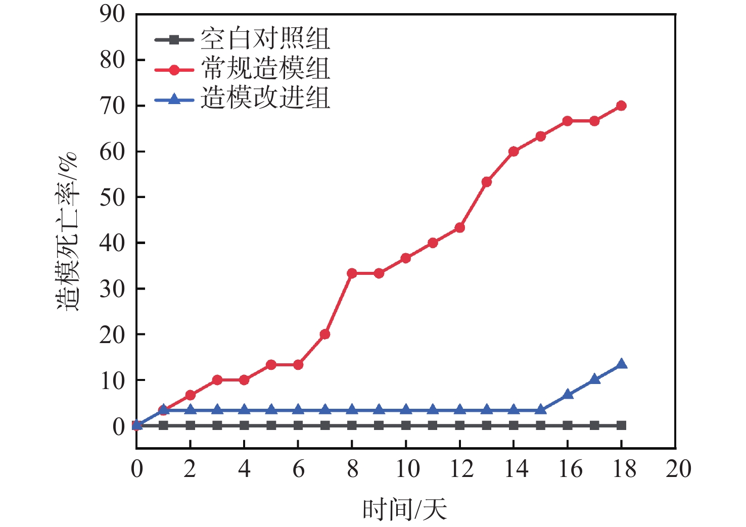
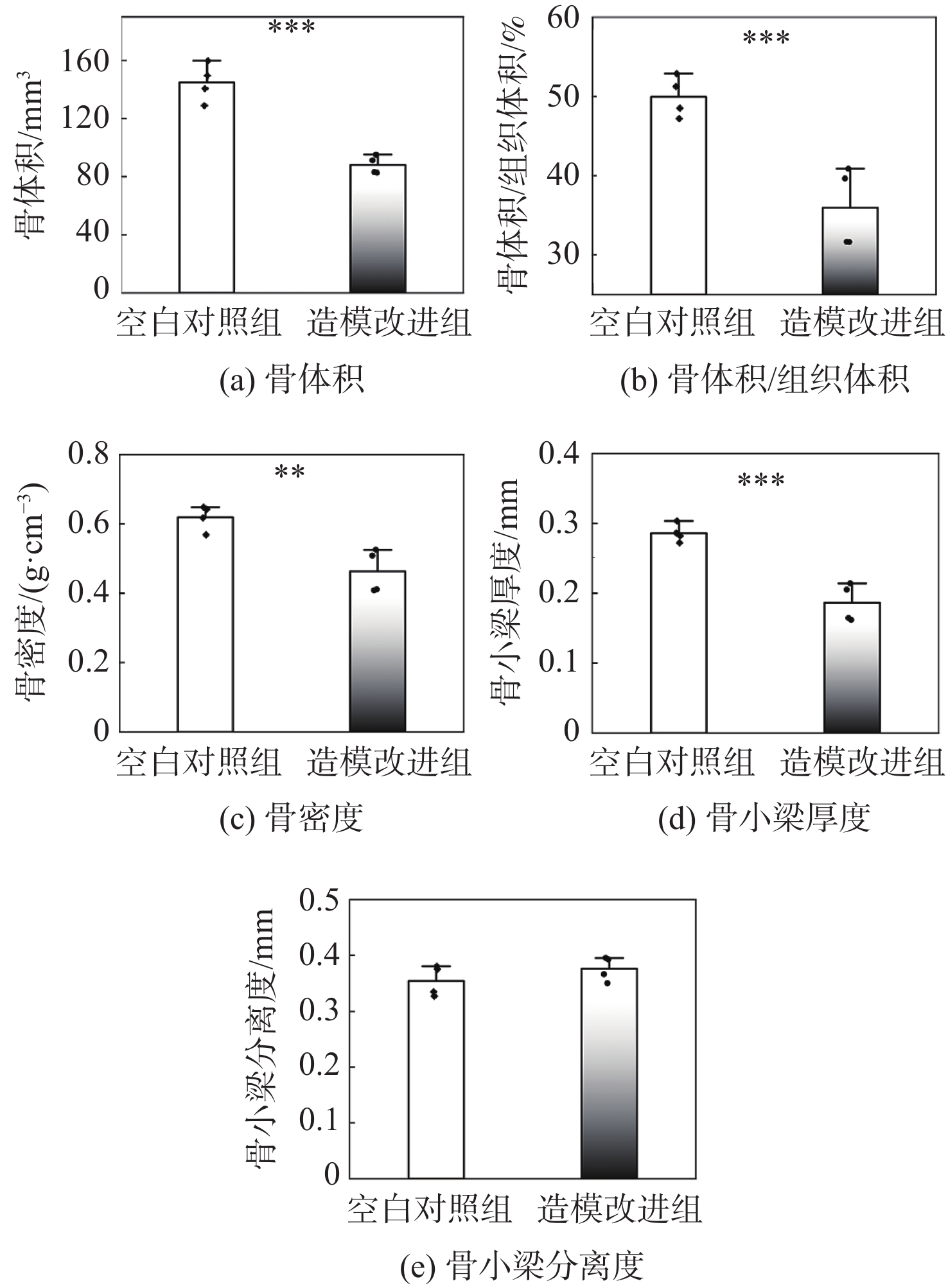
股骨头坏死动物模型的建立对临床上股骨头坏死的治疗具有重要意义。对目前最常见的兔激素性股骨头坏死的造模方法进行改进,并对改进后的造模结果进行评估,希望能够在降低造模动物死亡率的同时成功诱导股骨头坏死。选择周龄介于16~20周,体重介于2.8~3.2 kg的雄性新西兰大白兔90只,随机分为空白对照组、常规造模组和造模改进组3组。其中,空白对照组仅注射生理盐水;常规造模组在特定时间点注射脂多糖和甲基泼尼松龙;造模改进组在常规造模组的基础上辅以青霉素、雷尼替丁、地克珠利、肠道润滑剂和乳酶生干预。结果表明:造模改进组动物的死亡率(13.3%)远低于常规造模组(70%)。造模完成后的微型计算机断层扫描技术(Micro-CT)分析和组织学染色结果显示:造模改进组股骨头区域出现了早期股骨头坏死的特征,其骨体积、骨体积与组织体积比、骨密度、骨小梁厚度均显著低于空白对照组,而空骨陷窝个数相较于空白对照组则出现了显著提升,证明改进后的造模方法在降低死亡率的同时成功诱导了兔股骨头坏死。
-
关键词:
- 股骨头坏死 /
- 激素 /
- 动物模型 /
- 微型计算机断层扫描技术 /
- 组织学染色
Abstract:The establishment of animal model of femoral head necrosis is of great significance for the clinical treatment of this disease. To successfully induce femoral head necrosis while reducing the mortality of modeling animals, this study improves the commonly used modeling method of steroid-induced femoral head necrosis in rabbits. Ninety male New Zealand white rabbits (2.8~3.2 kg) are randomly divided into control group, conventional model group and improved model group. The control group is only injected with normal saline; the conventional model group is injected with lipopolysaccharide and methylprednisolone; the improved model group is supplemented with penicillin, ranitidine, diclazuril, intestinal lubricant and lactasin based on conventional model. The results show that the death rate of the improved model group (13.3%) is much lower than that of the conventional model group (70%). The Micro-CT and histological results exhibit that the bone volume, bone volume/tissue volume ratio, bone mineral density and trabecular thickness in improved model group are significantly lower than those in control group, while the number of empty bone lacunae is significantly higher than that in control group. These results demonstrate the successful establishment of steroid-induced femoral head necrosis in rabbits.
-
Key words:
- femoral head necrosis /
- steroid /
- animal model /
- Micro-CT /
- histological staining
-

图 1 激素性股骨头坏死动物模型造模过程中死亡因素
Figure 1. Death factors in animal model of steroid-induced femoral head necrosis

图 3 股骨头部位Micro-CT断层扫描及三维重建图像
Figure 3. Micro-CT tomography and 3D reconstruction images of femoral head

图 4 股骨头部位Micro-CT数据定量分析结果
Figure 4. Quantitative analysis of Micro-CT data in femoral head region
-
[1] WANG C K, HO M L, WANG G J, et al. Controlled-release of rhBMP-2 carriers in the regeneration of osteonecrotic bone[J]. Biomaterials, 2009, 30(25): 4178-4186. doi: 10.1016/j.biomaterials.2009.04.029 [2] MARUYAMA M, NABESHIMA A, PAN C C, et al. The effects of a functionally-graded scaffold and bone marrow-derived mononuclear cells on steroid-induced femoral head osteonecrosis[J]. Biomaterials, 2018, 187: 39-46. doi: 10.1016/j.biomaterials.2018.09.030 [3] ZHANG H X, ZHANG X P, XIAO G Y, et al. In vitro and in vivo evaluation of calcium phosphate composite scaffolds containing BMP-VEGF loaded PLGA microspheres for the treatment of avascular necrosis of the femoral head[J]. Materials Science and Engineering:C, 2016, 60: 298-307. doi: 10.1016/j.msec.2015.11.055 [4] WAEWSAWANGWONG W, RUCHIWIT P, HUDDLESTON J I, et al. Hip arthroplasty for treatment of advanced osteonecrosis: Comprehensive review of implant options, outcomes and complications[J]. Orthopedic Research and Reviews, 2016, 8: 13-29. doi: 10.2147/ORR.S35547 [5] STEVENS K, TAO C, LEE S U, et al. Subchondral fractures in osteonecrosis of the femoral head: Comparison of radiography, CT, and MR imaging[J]. American Journal of Roentgenology, 2003, 180(2): 363-368. doi: 10.2214/ajr.180.2.1800363 [6] QIN L, ZHANG G, SHENG H, et al. Multiple bioimaging modalities in evaluation of an experimental osteonecrosis induced by a combination of lipopolysaccharide and methylprednisolone[J]. Bone, 2006, 39(4): 863-871. doi: 10.1016/j.bone.2006.04.018 [7] 孙磊, 袁源, 牛睿, 等. 辐照和EO灭菌对SIS材料免疫原性的对比研究[J]. 北京航空航天大学学报, 2020, 46(12): 2245-2252. doi: 10.13700/j.bh.1001-5965.2019.0621SUN L, YUAN Y, NIU R, et al. Comparative study on immunoreactions of small intestinal submucosa by irradiation and ethylene oxide sterilization treatments[J]. Journal of Beijing University of Aeronautics and Astronautics, 2020, 46(12): 2245-2252(in Chinese). doi: 10.13700/j.bh.1001-5965.2019.0621 [8] 张林, 孙磊, 徐梦浛, 等. 可吸收胶原膜的体内免疫反应评价[J]. 北京航空航天大学学报, 2018, 44(4): 879-886. doi: 10.13700/j.bh.1001-5965.2017.0230ZHANG L, SUN L, XU M H, et al. Immunological response evaluation of absorbable collagen membrane in vivo[J]. Journal of Beijing University of Aeronautics and Astronautics, 2018, 44(4): 879-886(in Chinese). doi: 10.13700/j.bh.1001-5965.2017.0230 [9] XU J Z, GONG H P, LU S T, et al. Animal models of steroid-induced osteonecrosis of the femoral head-a comprehensive research review up to 2018[J]. International Orthopaedics, 2018, 42(7): 1729-1737. doi: 10.1007/s00264-018-3956-1 [10] 陈冠儒, 王萧枫, 王利明, 等. 兔激素性股骨头坏死造模中的致死因素与干预分析[J]. 浙江中西医结合杂志, 2012, 22(5): 342-344. doi: 10.3969/j.issn.1005-4561.2012.05.004CHEN G R, WANG X F, WANG L M, et al. Analysis on fetal factors and intervention of steriod-induced femoral head necrosis in rabbits[J]. Zhejiang Journal of Integrated Traditional Chinese and Western Medicine, 2012, 22(5): 342-344(in Chinese). doi: 10.3969/j.issn.1005-4561.2012.05.004 [11] 王程, 苟文隆, 徐小龙, 等. 人股骨头坏死标本不同区域骨小梁的显微结构特征及病理学表现[J]. 解放军医学院学报, 2014, 35(5): 463-465. doi: 10.3969/j.issn.2095-5227.2014.05.018WANG C, GOU W L, XU X L, et al. Microstructure features and pathology of trabecula in different regions of femoral head necrosis[J]. Academic Journal of Chinese PLA Medical School, 2014, 35(5): 463-465(in Chinese). doi: 10.3969/j.issn.2095-5227.2014.05.018 [12] HAMBLI R. Micro-CT finite element model and experimental validation of trabecular bone damage and fracture[J]. Bone, 2013, 56(2): 363-374. doi: 10.1016/j.bone.2013.06.028 [13] ZHANG G, SHENG H, HE Y X, et al. Continuous occurrence of both insufficient neovascularization and elevated vascular permeability in rabbit proximal femur during inadequate repair of steroid-associated osteonecrotic lesions[J]. Arthritis and Rheumatism, 2009, 60(10): 2966-2977. doi: 10.1002/art.24847 [14] LAI Y X, LI Y, CAO H J, et al. Osteogenic magnesium incorporated into PLGA/TCP porous scaffold by 3D printing for repairing challenging bone defect[J]. Biomaterials, 2019, 197: 207-219. doi: 10.1016/j.biomaterials.2019.01.013 [15] HA Y C, KIM H J, KIM S Y, et al. Effects of age and body mass index on the results of transtrochanteric rotational osteotomy for femoral head osteonecrosis: Surgical technique[J]. The Journal of Bone and Joint Surgery American Volume, 2011, 93(S1): 75-84. [16] JIN S Y, MENG C Q, HE Y, et al. Curcumin prevents osteocyte apoptosis by inhibiting M1-type macrophage polarization in mice model of glucocorticoid-associated osteonecrosis of the femoral head[J]. Journal of Orthopaedic Research, 2020, 38(9): 2020-2030. doi: 10.1002/jor.24619 [17] WU X, FENG X, HE Y, et al. IL-4 administration exerts preventive effects via suppression of underlying inflammation and TNF-alpha-induced apoptosis in steroid-induced osteonecrosis[J]. Osteoporosis International, 2016, 27(5): 1827-1837. doi: 10.1007/s00198-015-3474-6 [18] WANG C Q, XU H X, LIU C, et al. CaO2/gelatin oxygen slow-releasing microspheres facilitate tissue engineering efficiency for the osteonecrosis of femoral head by enhancing the angiogenesis and survival of grafted bone marrow mesenchymal stem cells[J]. Biomaterials Science, 2021, 9(8): 3005-3018. doi: 10.1039/D0BM02071K -

 下载:
下载:


 点击查看大图

计量
- 文章访问数: 221
- HTML全文浏览量: 44
- PDF下载量: 18
- 被引次数: 0
